ABCDEs of Melanoma

Contributed by Dr. Lim Wenkie
Moles are commonly seen on our skin. It is a skin growth that results from the clustering of melanocytes (pigmented cells). Most moles are actually benign and have no severe risk or threat to our health. However, some moles can become cancerous. You should get your moles evaluated regularly by your dermatologist to make sure that it is not showing signs of turning malignant. If so, then your dermatologist would likely remove the mole in its entirety to eradicate the cancer and prevent its spread.
So how does your friendly local dermatologist determine whether a mole is cancerous? As a general rule of thumb, medical professionals will follow the ABCDE visual cues.
Let’s go through the five signs of ABCDEs that would help determine whether a mole requires removal or not.
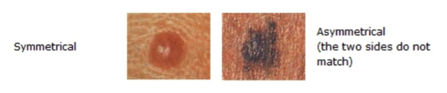
1. Asymmetrical Shape
Even though no mole is perfectly symmetrical, most benign moles are round and resembles a circle or an oval. If you were to draw an imaginary line going through the middle of a mole, basically both sides should more or less mirror each other. Moles that lacks this asymmetry might indicate irregular cellular activity, which may alert your doctor to think of skin cancer. After careful examination, your dermatologist may conduct a mole biopsy (removing a portion or the entire mole) to be further examine it for cancer. If the result comes back positive, they will recommend removing it entirely.
2. Borders that are ill-defined or irregular
Benign moles are also typically having well defined borders. On the other hand, malignant moles may have borders that are undefined and/or oddly shaped. Irregular borders may not always indicate skin cancer, but they will definitely prompt your dermatologist to have a closer look and also recommend more regular follow ups.

3. Colour and Consistency
Moles usually have different shades of brown, however it all depends on several factors. These factors include one’s skin type and skin tone, and also the amount of clustered pigment cells within the mole. However, while one mole might differ in colour from another, the colour of the mole should be of a consistent colour throughout the mole. If a single mole is darker in some areas yet lighter in others, this suggests an irregularity that your dermatologist will examine further during your mole evaluation.

4. Diameter of the mole
Just as a mole varies in shapes and colour, they also vary in size. Most benign moles are relatively small (less than 6 mm in diameter) in size. There are exceptions, of course. Even still, if a mole is larger than 6 mm, your dermatologist will check for potential of cancer and may suggest for removal of it.

5. Evolution of moles
Lastly, your doctor will take note of any changes in your moles, including its shape, colour, and even the appearance of new moles. We may experience new moles throughout our lives, though it commonly stops after adolescence, while some may even disappear on their own. New moles may be benign, especially if they are regular in shape, colour and size, but they are still worth getting it checked out. But if any of your existing moles have significantly changed in shape, colour and size, your dermatologist might conduct further tests and may recommend removal of them.

The ABCDEs of melanoma are important during a mole check-up. But of course, these aren’t the only reasons your might have a mole removal. You might simply not like the appearance and/or the location of a benign mole and ask your dermatologist to remove it for cosmetic purposes. So, make sure your check with your dermatologist if you have any doubts and concerns about your mole.
Reference:
- John Breneman ― Towards Early-Stage Malignant Melanoma Detection Using Consumer Mobile Devices‖ Stanford Center for Professional Development, 2012
- National cancer institute, what you need to know about melanoma other skin cancers. Visit us at http://www.cancer.gov
- Melanoma Symptoms, [Online]. Available: http://www.cancerhelp.org/ help/default.asp?page=3009, National Institute for Health and Clinical Exce-lence, Tech. Rep.
- Gambichler, P. Regeniter, F. Bechara, A. Orlikov, R. Vasa, G. Moussa, M. Stücker, P. Altmeyer, and K. Hoffmann, ―Characterization of benign and malignant melanocytic skin lesions using optical coherence tomography in vivo,‖ Journal of the American Academy of Dermatology, vol. 57, no. 4, pp. 629–637, 2007.
- Unsupervised segmentation of skin lesions – Research Gate. Available from: http://www.researchgate.net/publication/33418237_Unsupervised_segmentation_of_ skin_lesions [accessed Nov 16, 2015]
